Communication

Developing an Enzyme-Assisted Derivatization Method for Analysis of C27 Bile Alcohols and Acids by Electrospray Ionization-Mass Spectrometry

Swansea University Medical School, ILS1 Building, Swansea University, Singleton Park, Swansea SA2 8PP, Wales, UK
*
Author to whom correspondence should be addressed.
Molecules 2019, 24(3), 597; https://doi.org/10.3390/molecules24030597
Submission received: 21 December 2018 / Revised: 29 January 2019 / Accepted: 30 January 2019 / Published: 7 February 2019

Abstract

:
Enzyme-assisted derivatization for sterol analysis (EADSA) is a technology designed to enhance sensitivity and specificity for sterol analysis using electrospray ionization–mass spectrometry. To date it has only been exploited on sterols with a 3β-hydroxy-5-ene or 3β-hydroxy-5α-hydrogen structure, using bacterial cholesterol oxidase enzyme to convert the 3β-hydroxy group to a 3-oxo group for subsequent derivatization with the positively charged Girard hydrazine reagents, or on substrates with a native oxo group. Here we describe an extension of the technology by substituting 3α-hydroxysteroid dehydrogenase (3α-HSD) for cholesterol oxidase, making the method applicable to sterols with a 3α-hydroxy-5β-hydrogen structure. The 3α-HSD enzyme works efficiently on bile alcohols and bile acids with this stereochemistry. However, as found by others, derivatization of the resultant 3-oxo group with a hydrazine reagent does not go to completion in the absence of a conjugating double bond in the sterol structure. Nevertheless, Girard P derivatives of bile alcohols and C27 acids give an intense molecular ion ([M]+) upon electrospray ionization and informative fragmentation spectra. The method shows promise for analysis of bile alcohols and 3α-hydroxy-5β-C27-acids, enhancing the range of sterols that can be analyzed at high sensitivity in sterolomic studies.

Graphical Abstract

1. Introduction

Sterols represent one of the major classes of lipids found in living systems [1]. In mammals, cholesterol represents the archetypal sterol. It is metabolized through a myriad of intermediates to C21–18 steroids and to C24 bile acids [2,3,4,5,6,7,8,9]. For decades there was little interest in these intermediates, however, in recent years the situation has changed with the realization that intermediates in bile acid biosynthesis are ligands to nuclear receptors, including the liver X receptors (LXRs, NR1H3, NR1H2) [10,11,12,13], farnesoid X receptor (FXR, NR1H4) [14], pregnane X receptor (PXR, also known as xenobiotic sensing nuclear receptor, SXR, NR1I2) [15,16], RAR-related orphan receptor γt (RORγt, NR1F3) [17], and estrogen receptors (ERs, NR3A1, NR3A2) [18]. They are also related to G protein-coupled receptors (e.g., Epstein-Barr virus induced gene 2 (EBI2, GPR183) [19,20] and smoothened (SMO, FZD11) [21,22]), and are involved in the regulation of cholesterol biosynthesis by binding to INSIG1 (insulin induced gene 1) [23]. Cholesterol metabolites have traditionally been analyzed by gas-chromatography-mass spectrometry (GC-MS) [3,4,6,24,25], however, liquid chromatography (LC)-MS, is currently taking a dominant role in their analysis [5,26].
Analysis of cholesterol metabolites is valuable for the diagnosis of rare inborn errors of metabolism, and defects in bile acid and steroid synthesizing enzymes are efficiently characterized by LC-MS or GC-MS analysis of plasma or urine [6,27,28,29]. By performing these analyses, unexpected metabolites are identified, which are normally only minor components of the sterolome, but which are abundant in the disease state [7,8,9]. When these unexpected metabolites are considered, it becomes evident that the complexity of the sterolome is enormous. Sterolomics is one of the subdivisions of lipidomics, however, in most lipidomic studies, sterols are underrepresented; this is because, other than cholesterol and its esters, they are not abundant, and their ionization characteristics in positive-ion electrospray ionization (ESI)-MS (the dominant ionization method in lipidomics) are poor [30,31]. To improve the sensitivity for sterol analysis, many groups adopt a derivatization strategy where sterols are chemically modified to improve their ionization characteristics [32,33,34,35,36,37,38,39].
One derivatization strategy that has lately become popular is enzyme-assisted derivatization for sterol analysis (EADSA, Scheme 1) [8,9,12,13,17,40,41,42,43,44,45,46,47]. EADSA technology was designed to add specificity and sensitivity to sterol analysis [40,44,45,46]. This is achieved by specifically targeting the 3β-hydroxy-5-ene or 3β-hydroxy-5α-hydrogen function in sterols and converting the 3β-hydroxy to a 3-oxo group with bacterial cholesterol oxidase from Brevibacterium or Streptomyces sp. [40,41]. Once introduced, the 3-oxo group is derivatized with the positively charged Girard hydrazine reagent, introducing a charge-tag to the target analyte and improving sensitivity in ESI-MS. A limitation of the existing protocol is that it is not applicable to sterols with a 3α-hydroxy group. Here we describe how this limitation is overcome for the analysis of bile alcohols and C27 bile acids with this stereochemistry. The methodology is potentially applicable for C24 bile acids but requires further optimization to achieve similar sensitivity as for C27 alcohols and acids.

2. Results

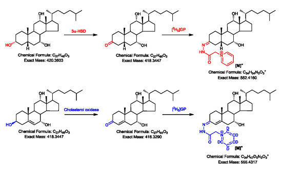
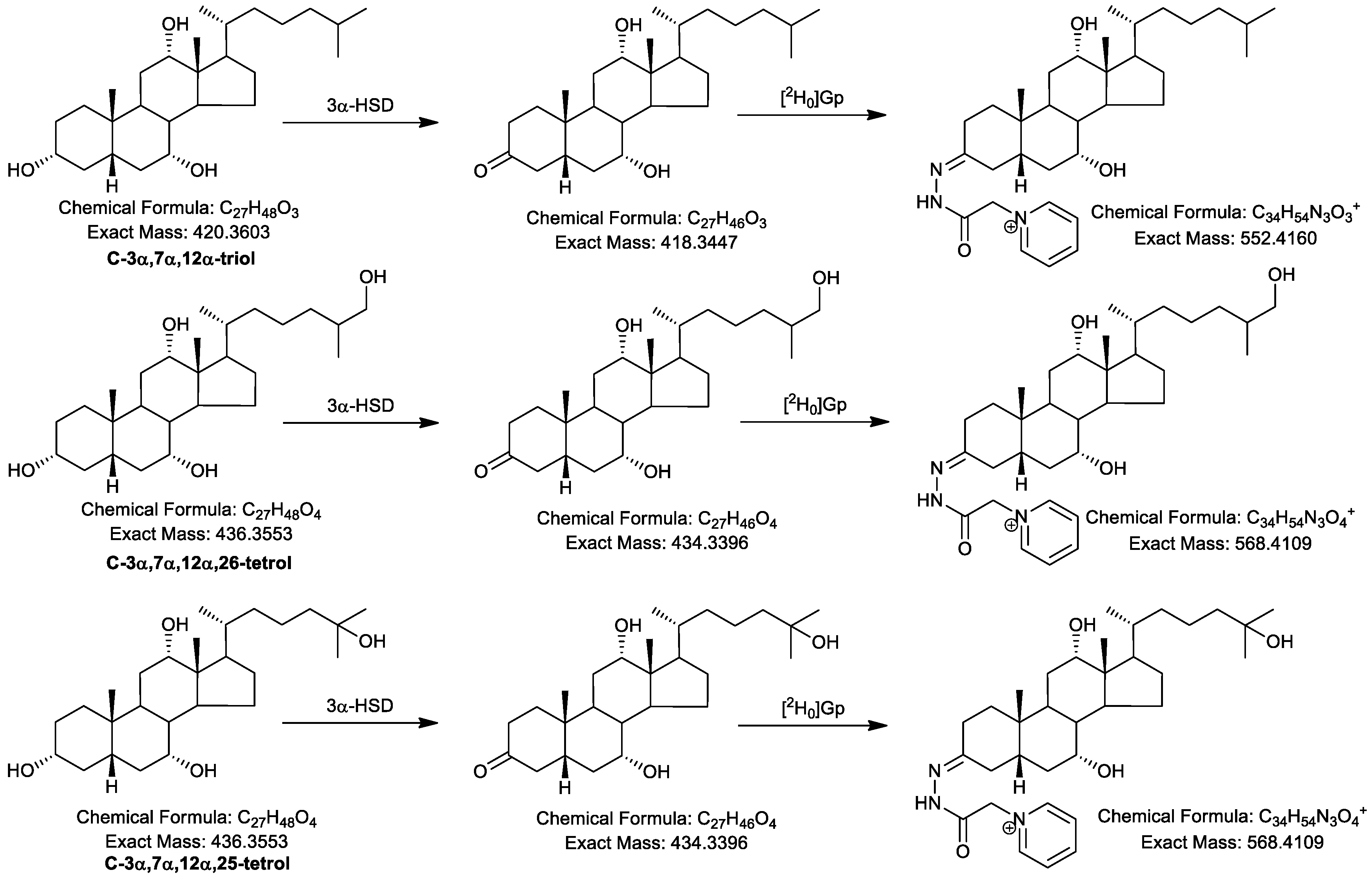
2.1. Oxidation Efficiency of 3α-Hydroxysteroid Dehydrogenase (3α-HSD)

The efficiency of oxidation of 3α-HSD towards 3α-hydroxy-5β-substrates was evaluated using cholic acid (3α,7α,12α-trihydroxy-5β-cholan-24-oic acid, BA-3α,7α,12α-triol) and 3α,7α,12α-trihydroxy-5β-cholestan-26-oic acid (CA-3α,7α,12α-triol) as representative analytes, because unlike neutral sterols, C24 and C27 bile acids are readily ionized by ESI (negative-ion mode) and can be detected in LC-MS analysis in both their 3α-hydroxy or 3-oxo forms. To determine oxidization efficiency, the amounts of unoxidized (3α-hydroxy) and oxidized (3-oxo) acids were determined after different incubation periods and incorporated into Equation (1).
Oxidation efficiency (%) = amount oxidized/(amount oxidized + amount unoxidized) × 100%
Using similar reaction conditions to those employed in the current study (see Section 4.2.1), Une et al. have shown that an incubation time of 20 h leads to about 95% conversion of the 3α-hydroxy group to the 3-one in cholic acid and in most bile alcohols [48]. We found that reducing the incubation time to 14 h gave a similarly high yield (>95%) of product for CA-3α,7α,12α-triol and cholic acid. Either incubation time, 14 h or 20 h, gave satisfactory results. In the current study, similarly high efficiencies of oxidation (>95%) were found for both the glycine and taurine conjugates of cholic acid and for unconjugated chenodeoxycholic (3α,7α-dihydroxy-5β-cholan-24-oic), deoxycholic (3α,12α-dihydroxy-5β-cholan-24-oic) and lithocholic (3α-hydroxy-5β-cholan-24-oic) acids.

2.2. Girard P (GP)-Derivatisation of 3-Oxo Groups

Previous studies have shown that hydrazone formation is very efficient towards 3-oxo-4-ene substrates and to other α,β-unsaturated ketones [40,41,49,50]. In earlier EADSA studies using cholesterol oxidase in phosphate buffer to convert 3β-hydroxy-5-ene sterols to their 3-oxo-4-ene equivalents, GP derivatization was achieved by simply adding methanol to the incubation solution to give a 70% methanol solution and then adding acetic acid and GP hydrazine reagent [40,41,45,46,50,51,52]. However, the buffer required for 3α-HSD oxidation of the 3α-hydroxy group to the 3-one is 100 mM pyrophosphate buffer, pH 8.9, and upon methanol addition, necessary for subsequent hydrazone formation, a precipitate is formed. This can be avoided by limiting the methanol content to 5%, however, under these conditions hydrazone formation is reversed back to the hydrazine and free carbonyl. For this reason, following incubation with 3α-HSD, samples were desalted on an Oasis HLB reversed-phase column and eluted in methanol, a solvent suitable for subsequent GP hydrazone formation. The GP derivatization efficiency was assessed by LC-MS in the negative-ion mode by comparing the amount of underivatized oxidized acid present before and after the GP derivatization step and incorporating the data into Equation (2).
Derivatization efficiency (%) = 100% − [(amount underivatized acid after derivatization/amount underivatized acid before derivatization) × 100%]
Unlike 3-oxo-4-ene sterols, which are derivatized with 100% efficiency in acidic methanol [40], the derivatization efficiency for the 3-oxo-5β-hydrogen compounds formed from their 3α-hydroxy-5β-hydrogen substrates (cholic, chenodeoxycholic, deoxycholic acids and CA-3α,7α,12α-triol), was only 45–60%. Taurine- and glycine-conjugated cholic acid gave a similar degree of derivatization efficiency after 3α-HSD oxidation. Despite the moderate yield of derivatization products, the high sensitivity provided by GP derivatization of unconjugated substrates (see Section 2.3.) negates this imperfection.

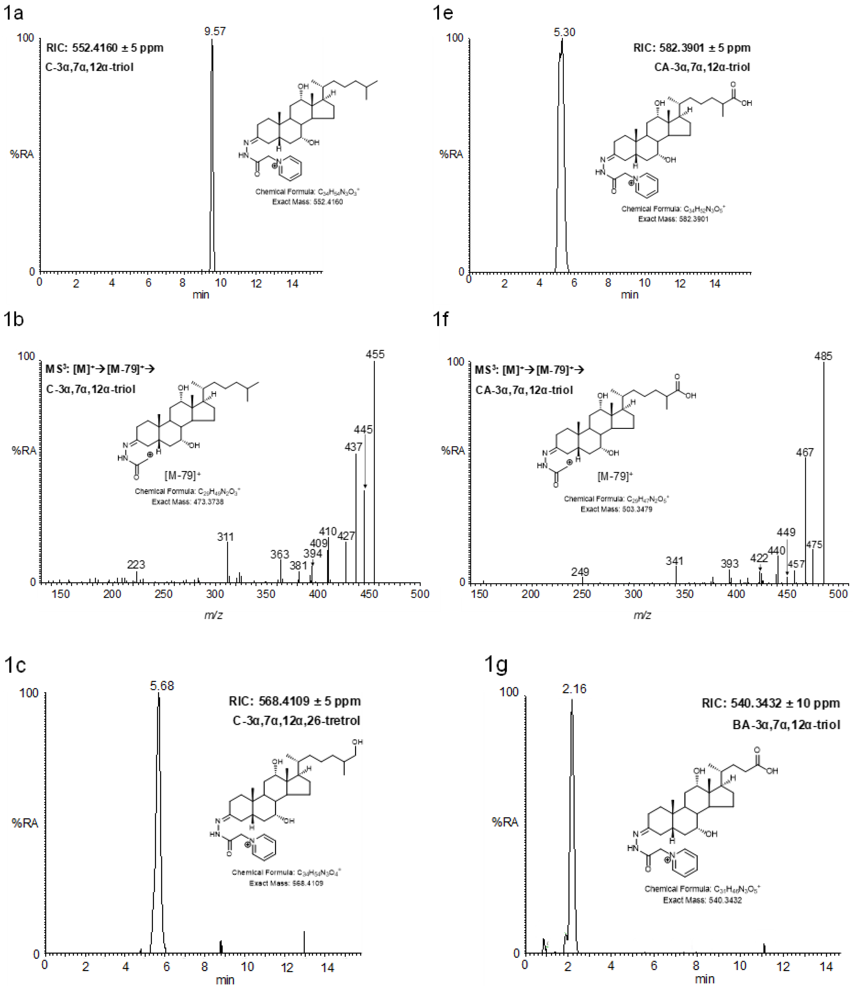
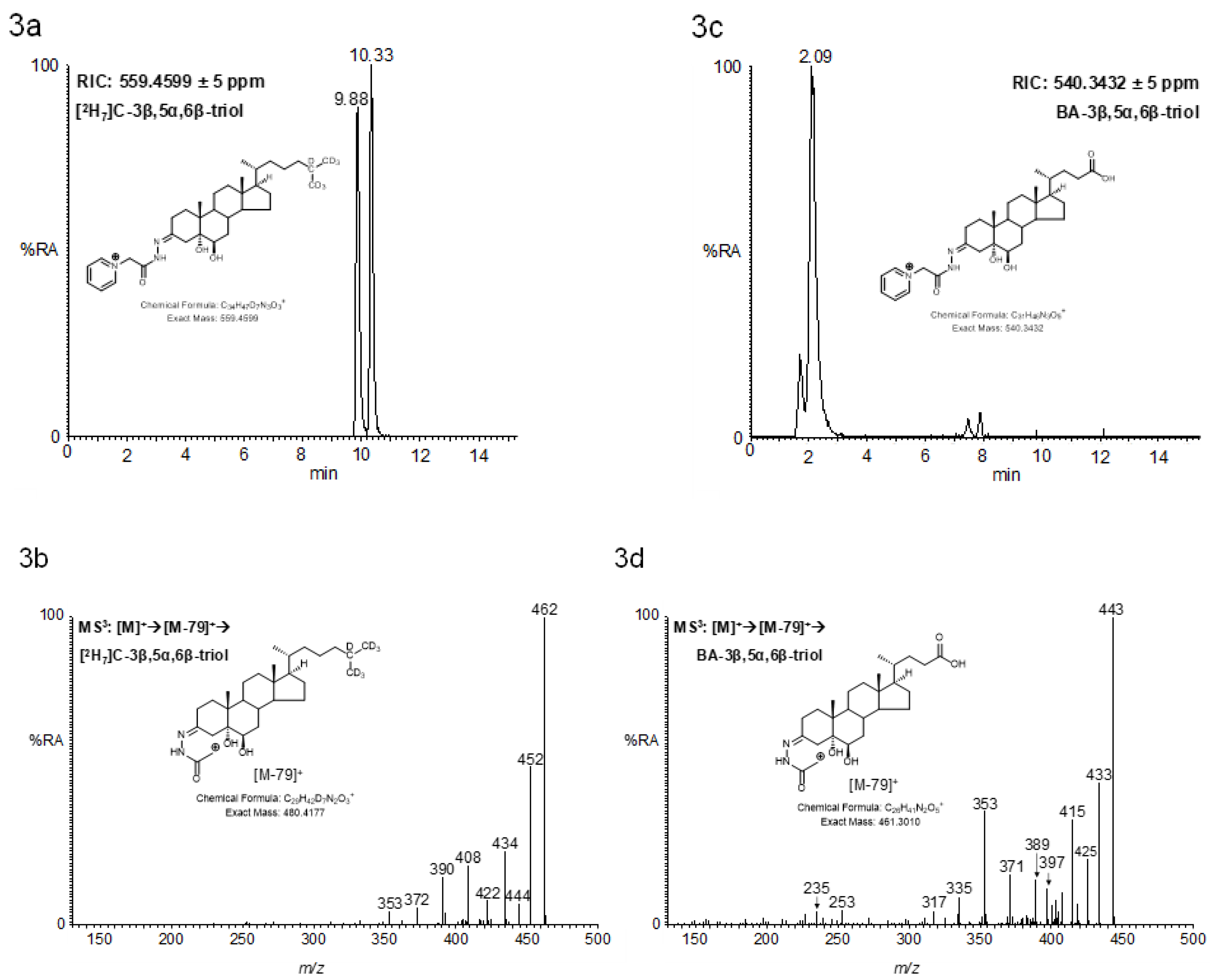
2.3. LC-MS Analysis of Oxidised/Derivatised 3α-Hydroxy-5β-Hydrogen Substrates

In this preliminary study we have not optimized the chromatographic or MS conditions for the GP-derivatized target compounds but rather used existing LC-MS conditions used previously for GP-derivatized sterols [44,45,46,52]. The logic behind this is that by using isotope-labelled GP reagent, the ultimate aim will be to analyze sterols oxidized with cholesterol oxidase or 3α-HSD in a single LC-MS run. Neither have we performed detailed investigations of limits of quantification or linearity of dynamic range in the current study. However, we find that the sensitivity obtained here for 3α-HSD oxidized/GP-derivatized C27 sterols with a 3α-hydroxy-5β-stereochemistry is of the same order of magnitude to that obtained for GP derivatives generated after cholesterol oxidase treatment of 3β-hydroxy-5-ene substrates. For the C27 substrates an on-column limit of detection (LOD, signal/noise, 5:1) of 250 fg was achieved. The on-column LOD for the C24 acid, cholic acid, was 250 pg. More work is required to explain this discrepancy in sensitivity and the even poorer sensitivity with glycine- and taurine-conjugated acids. Optimization of the ion-source conditions for different groups of analytes, or at least compromise in the settings chosen, is likely to be necessary.

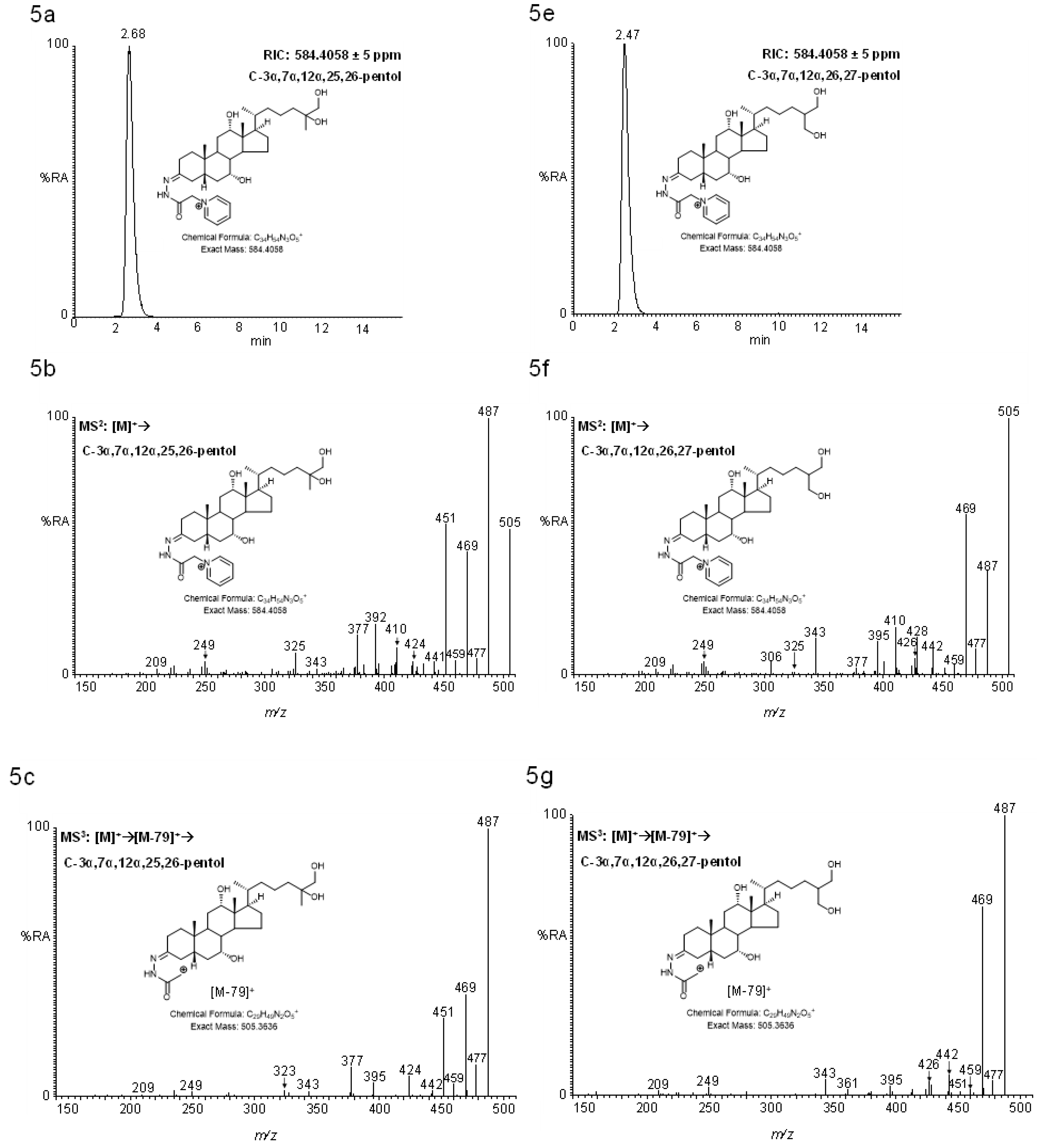
2.4. MSn Fragmentation

A major driver for the current study was the poor fragmentation properties of unconjugated C24 and C27 bile acids under conditions of ESI–tandem MS (MS/MS) at low collision energy (<100 eV) [5,53,54,55,56] (see also MassBank of North America http://mona.fiehnlab.ucdavis.edu/). This has led to many studies in which the precursor ion at unit mass resolution is also used as the “product ion” for generation of LC–multiple reaction monitoring (MRM) chromatograms. Once derivatized with the GP reagent, both bile acids and bile alcohols fragment under low-energy conditions with the loss of the pyridine group, resulting in [M-Py]+ ions (see Scheme 1). These ions can be fragmented further in ion-trap instruments to give multistage fragmentation (MS3, [M]+→[M-Py]+→) spectra rich in fragment ions. The advantage of MS3 is that it provides an extra dimension of separation compared to MS2, where spectra are a composite of fragment ions derived from desired and undesired coselected precursor ions.

2.4.1. Triols and Tetrols
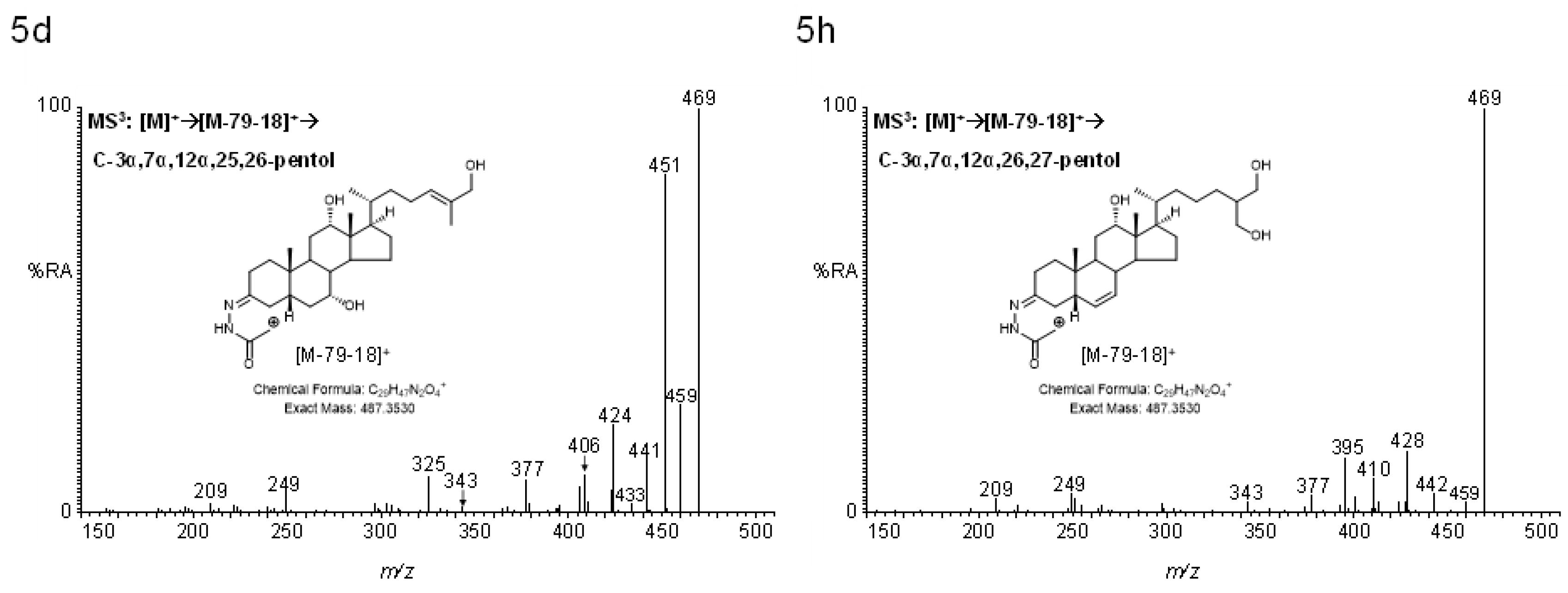
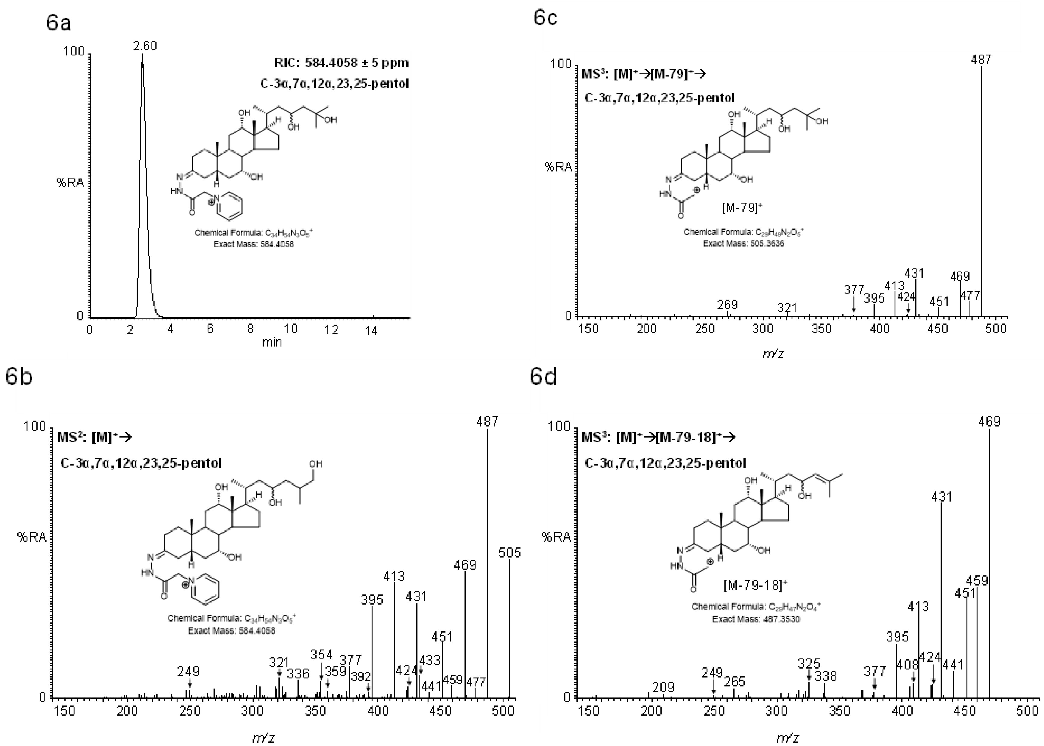
Shown in Figure 1 are representative reconstructed-ion chromatograms (RICs) and MS3 ([M]+→[M-Py]+→) spectra of oxidized/GP-derivatized C27 bile alcohols 5β-cholestane-3α,7α,12α-triol (C-3α,7α,12α-triol) and 5β-cholestane-3α,7α,12α,26-tetrol (C-3α,7α,12α,26-tetrol)), the C27 acid (CA-3α,7α,12α-triol), and the C24 trihydroxy bile acid (cholic acid).
The MS3 spectra show considerable similarity, with many fragment ions in the spectrum of the C26 acid and tetrol being shifted by m/z 30 and m/z 16, respectively from the corresponding triol. This is explained by the introduction of a carboxylic acid group (+ O2 − H2) or hydroxy (+ O) group to the terminal carbon (C-26) of the sterol side-chain (Scheme 2). Postulated structures of fragment ions for C-3α,7α,12α-triol are shown in Scheme 3 and for C-3α,7α,12α,26-tetrol and CA-3α,7α,12α-triol in Supplemental Schemes S1 and S2, and are listed in Table 1.
When a hydroxy group is positioned at C-25 rather than C-26, the lability of the hydroxy group leads to a more intense [M-Py-18]+ than [M-Py]+ ion in the MS2 ([M]+→) spectrum of 5β-cholestane-3α,7α,12α,25-tetrol (C-3α,7α,12α,25-tetrol, Figure 2b) than in its epimer C-3α,7α,12α,26-tetrol (Supplemental Figure S1b). See Table 1 to correlate m/z with fragment ion composition. The MS3 ([M]+→[M-Py-18]+→) spectrum of C-3α,7α,12α,25-tetrol (Figure 2d) is almost identical to the MS3 ([M]+→[M-Py]+→) of C-3α,7α,12α-triol (Figure 1b) but with an offset of m/z -2 (+ O – H2O, see also Supplemental Scheme S3).
Unsurprisingly, the MS3 ([M+→[M-Py]+→) spectrum of the C24 acid, cholic acid (Figure 1h), shows the same pattern of fragment ions as the C27 acid CA-3α,7α,12α-triol (Figure 1f) but offset by m/z -42 (-C3H6), corresponding to the mass difference between equivalent C27 and C24 acids (cf. Supplemental Schemes S2 and S4).
A key structurally distinct fragment ion for all the 3α,7α,12α-triols is the [A3-H-(H2O)2]+ ion (or [A3-H-(H2O)3]+ when an additional hydroxy group is at C-25, Table 1), a triply unsaturated carbonium ion consisting of B-, C- and D-rings plus the C17 side-chain, where charge is delocalized across the three double bonds in the ring system (Scheme 3, inset). An equivalent fragment ion is not observed in cholesterol oxidase-oxidized/GP-derivatized 3β,5α,6β-triols (Figure 3).

2.4.2. Pentols

The human autosomal recessive disease, cerebrotendinous xanthomatosis (CTX), results from a deficiency in cytochrome P450 27A1 (CYP27A1) [57], a key enzyme in the conversion of cholesterol to bile acids [28,29]. As a consequence of this deficiency, polyhydroxy-bile alcohols are produced [58,59], providing an alternative route for bile acid biosynthesis and cholesterol removal [60].
As with C-3α,7α,12α,25-tetrol, the presence of a labile 25-hydroxy group in the epimers 5β-cholestane-3α,7α,12α,24R,25-pentol (C-3α,7α,12α,24R,25-pentol) and 5β-cholestane-3α,7α,12α,24S,25-pentol (C-3α,7α,12α,24S,25-pentol) results in abundant [M-Py-18]+ ions in the MS2 ([M]+→) spectra, and the MS3 ([M]+→[M-Py-18]+→) spectra resembles the MS3 ([M]+→[M-Py]+→) spectra of cholestanetetrols but is offset by m/z -2 (+ O – H2O) (Figure 4). Although the MSn spectra are very similar, the two epimers are readily separated on the LC column. Here in the MS2 ([M]+→) and MS3 ([M]+→[M-Py]+) spectra, in addition to the [A3-H-(H2O)2]+ fragment ion, a [A3-H-(H2O)3]+ fragment ion is also prominent (See Table 1 to correlate m/z with fragment ion composition).
Movement of the hydroxy group from C-24 to C-26 as in 5β-cholestane-3α,7α,12α,25,26-pentol (C-3α,7α,12α,25,26-pentol) results in a small delay in retention time and subtle changes to the MS2 ([M]+→) and MS3 ([M]+→[M-Py]+→ and [M]+→[M-Py-18]+→) spectra, for example, reduced abundance of fragment ions having lost four water molecules compared the equivalent having lost three water molecules (i.e., [M-79-36-36]+/[M-79-36-18]+) (Figure 5b–d, cf. Figure 4b–d,f–h). The bile alcohol 5β-cholestane-3α,7α,12α,26,27-pentol (C-3α,7α,12α,26,27-pentol) elutes between the 24R- and 25S-epimers of C-3α,7α,12α,24,25-pentol, but gives very different MSn spectra to the other cholestanepentols on account of the absence of a labile C-25-hydroxy group (Figure 5f–h, Supplemental Scheme S6). This is reflected in the ratio of fragment ions [M-79-36-18]+/[M-79-36]+ which is greatly reduced compared to pentols with a 25-hydroxy group. The comparative stability of the primary hydroxy groups at the termini, C-26 and C-27, is further reflected by an absence of fragment ions having lost four water molecules (e.g., [M-79-36-36]+).
Although it co-elutes with C-3α,7α,12α,24S,25-pentol, 5β-cholestane-3α,7α,12α,23,25-pentol (C-3α,7α,12α,23,25-pentol) gives unique MSn spectra (Figure 6). Each of the MS2 ([M]+→), MS3 ([M]+→[M-Py]+→) and MS3 ([M]+→[M-Py-18]+→) spectra show an unusual pattern of fragment ions at m/z 431.3, 413.3, and 395.3. It is not immediately obvious why this triad of fragment ions is so distinct for this molecule. Neither are the structures or chemical compositions of all these fragments easy to reconcile with the MS3 ([M]+→[M-Py-18]+→) spectrum.

3. Discussion

In this communication we describe preliminary studies to develop an enzyme-assisted derivatization for C27 bile alcohols and acids with a 3α-hydroxy-5β-hydrogen stereochemistry. The method still requires further optimization, particularly with respect to the GP-derivatization step which only gave a 45%–60% yield. Despite this, the considerable sensitivity of GP-derivatives makes the moderate yield tolerable. Although the LC-MS sensitivity for detection of C24 acids was not as good as for C27 acids, the rich MS3 fragment ion spectra provide a significant advantage over conventional MS/MS spectra of unconjugated acids where few fragment ions are observed. The on-column detection limit of 250 fg for C27 analytes translates to a limit of detection of about 0.2 ng/mL if 100 µL of biological fluid is worked up and 1% injected on-column, as in our usual procedure with EADSA [52]. For comparison, Johnson et al. could measure CA-3α,7α,12α-triol, after derivatization to the dimethylaminoethyl ester, at a concentration of about 60 ng/mL in as little as 5 µL of plasma, with 20% injected-on column [38], while DeBarber et al. determined the limit of quantification of 7α,12α-dihydroxy-5β-cholestan-3-one, the 3-oxo form of C-3α,7α,12α-triol, to be 20 ng/mL from 4 µL of plasma after derivatization to the oxime with (O-(3-trimethylammoniumpropyl) hydroxylamine) bromide [39]. We have not yet rigorously tested the repeatability of the EADSA methodology in biological samples. This will become relevant with the availability of isotope-labelled standards, which can be synthesized by methods described by Johnson et al. and by Shoda et al. [61]. Isotope-labelled internal standards will similarly facilitate the progression of the method to a quantitative format. We did not attempt to optimize LC-MS conditions for the GP-derivatives analyzed in this study; instead we used previously optimized conditions for derivatized oxysterols. The logic behind this was to allow the expansion of our sterol profiling method to include bile acids and alcohols derivatized with [2H0]GP after 3α-HSD treatment and oxysterols, and cholestenoic and cholenoic acids derivatized with [2H5]GP after cholesterol oxidase treatment, or vice versa, in a single LC-MS run. At present there are challenges with this strategy, as efficient ionization of glycine- and taurine-conjugated bile acids requires different ion-source conditions from the unconjugated GP-derivatives.

4. Materials and Methods

4.1. Materials

CA-3α,7α,12α-triol (LMST04030001) and [2H7]C-3β,5α,6β-triol were from Avanti Polar Lipids (Alabaster, AL, USA). Bile alcohols, C-3α,7α,12α-triol (LMST04030035), C-3α,7α,12α,25-tetrol (LMST04030037) and C-3α,7α,12α,26-tetrol, (LMST04030159 or LMST04030160), C-3α,7α,12α,24R,25-pentol (LMST04030177), C-3α,7α,12α,24S,25-pentol (LMST04030039), C-3α,7α,12α,25,26-pentol (LMST04030016), 3α,7α,12α,26,27-pentol (LMST04030041) and C-3α,7α,12α,23,25-pentol (LMST01010240 or LMST01010241) were kind gifts from Professor Jan Sjӧvall, Karolinska Institutet, Stockholm. BA-3β,5α,6β-triol (LMST04010339) was a kind gift from Professor Douglas Covey, Washington University. Other C24 bile acids were from Sigma-Aldrich (Dorset, UK) or Fluka Chemie (Buchs, Switzerland). 3α-Hydroxysteroid dehydrogenase (3α-HSD) from Pseudomonas testosteroni was from Sigma-Aldrich (Dorset, UK). β-Nicotinamide adenine dinucleotide hydrate and sodium pyrophosphate decahydrate were from Sigma-Aldrich. [2H0]GP ([1-(carboxymethyl)pyridinium chloride hydrazide]) reagent was from TCI Europe (Oxford, UK). [2H5]GP reagent was synthesized as the bromide salt as described in Crick et al. [45]. Solid phase extraction (SPE) columns, certified Sep-Pak C18, 200 mg (3 cm3), and 60 mg Oasis HLB (3 cm3), were from Waters Inc. (Elstree, UK). Solvents were obtained from Fisher Scientific (Loughborough, UK). Acetic acid and formic acid were of AnalaR NORMAPUR grade (BDH, VWR, Lutterworth, UK).

4.2. Methods

4.2.1. Oxidation and Derivatization

Oxidation of analytes by 3α-HSD was essentially as described by Une et al. [48]. β-NAD+ hydrate (19.8 mg) was dissolved in 100 mM pyrophosphate buffer pH 8.9 (1 mL). Analyte (40–400 ng) dissolved in ethanol (10 µL) was added to the buffered solution giving a final concentration of 1% ethanol, followed by addition of 3α-HSD (0.06 units). After incubation at room temperature for 20 h, methanol (40 µL) was added (giving an organic content of 5%). To separate oxidized analyte from buffer, the solution was loaded onto a HLB column (60 mg, previously washed with methanol, 6 mL, and conditioned with 5% methanol, 6 mL) followed by a rinse with 5% methanol (0.5 mL). The column was then further washed with 5% methanol (6 mL). Analytes were eluted with methanol (2 mL). For samples to be analyzed by ESI in the negative-ion mode, to monitor oxidation efficiency, the methanol eluate was diluted with water to give a 60% methanol solution and was analyzed by LC-MS on the Orbitrap-Elite high resolution mass spectrometer (Thermo Fisher Scientific, Waltham, MA, USA) at 120,000 resolution (full width at half maximum height at m/z 400). To derivatize samples with GP reagent, glacial acetic acid (150 µL) was added followed by GP reagent (150 mg chloride salt, 190 mg bromide salt) and the mixture was left at room temperature overnight. The next day, water (1 mL) was added immediately prior to a second SPE step. This second SPE step was performed with recycling on an Oasis HLB column (60 mg) to remove excess derivatization reagent and was carried out as described in Abdel-Khalik et al. [52].

4.2.2. LC-MS(MSn) Analysis

LC-MS(MSn) was performed in the positive-ion mode as described in Abdel-Khalik et al. utilizing the Orbitrap-Elite hybrid MS preceded by a Dionex Ultimate 3000 LC system (Dionex, now Thermo Fisher Scientific) [52]. For analysis of underivatized acids in the negative-ion mode, other than for polarity reversal and a change of column from a Hypersil Gold C18 to a Kinetex core-shell technology XB-C18 column (2.6 µm, 2.1 mm × 50 mm, Phenomenex, Macclesfield, UK), the method was as for positive-ion mode LC-ESI-MS(MSn) as described in Abdel-Khalik et al. [52].

5. Patents

The derivatization method described in this manuscript is patented by Swansea University (US9851368B2) and licensed by Swansea Innovations to Avanti Polar Lipids and to Cayman Chemical Company.

Supplementary Materials

The following are available online. Supplemental Figure S1. MS2 ([M]+→) spectra of (a) C-3α,7α,12α-triol and (b) C-3α,7α,12α,26-tetrol, and (c) MS3 ([M]+→[M-Py-18]+→) spectrum of C-3α,7α,12α,26-tetrol. Data were generated on the Orbitrap-Elite mass spectrometer as in Figure 1. Supplemental Figure S2. Mass spectra of oxidized/GP-derivatized bile alcohols and acids recorded at the peak of their chromatographic elution. Except for C-3α,7α,12α,26-tetrol, C-3α,7α,12α,25,26-pentol, C-3α,7α,12α,26,27-pentol and C-3α,7α,12α,23,25-pentol which were injected (35 µL) on-column at 0.2 pg/µL all other analytes were inject on-column at 2 pg/µL. (a) C-3α,7α,12α-triol, (b) C-3α,7α,12α,26-tetrol, (c) CA-3α,7α,12α-triol, (d) C-3α,7α,12α,25-tetrol, (e) [2H7]C-3β,5α,6β-triol, (f) BA-3β,5α,6β-triol, (g) C-3α,7α,12α,24R,25-pentol, (h) C-3α,7α,12α,24S,25-pentol, (i) C-3α,7α,12α,25,26-pentol, (j) C-3α,7α,12α,26,27-pentol and (k) C-3α,7α,12α,23,25-pentol. Data were generated on the Orbitrap-Elite mass spectrometer as in Figure 1. Scheme S1: MS2 and MS3 fragmentation of oxidized/GP derivatized C-3α,7α,12α,26-tetrol. For simplicity the fragmented Girard derivatizing group is shown in its linear isomeric form. Cyclic forms are depicted in Scheme 1. Scheme S2: MS2 and MS3 fragmentation of oxidized/GP derivatized CA-3α,7α,12α-triol. For simplicity the fragmented Girard derivatizing group is shown in its linear isomeric form. Cyclic forms are depicted in Scheme 1. Scheme S3: MS2 and MS3 fragmentation of oxidized/GP derivatized C-3α,7α,12α,25-tetrol. For simplicity the fragmented Girard derivatizing group is shown in its linear isomeric form. Cyclic forms are depicted in Scheme 1. Scheme S4: MS2 and MS3 fragmentation of oxidized/GP derivatized BA-3α,7α,12α-triol. For simplicity the fragmented Girard derivatizing group is shown in its linear isomeric form. Cyclic forms are depicted in Scheme 1. Scheme S5: MS2 and MS3 fragmentation of oxidized/GP derivatized C-3α,7α,12α,25,26-pentol. For simplicity the fragmented Girard derivatizing group is shown in its linear isomeric form. Cyclic forms are depicted in Scheme 1. Scheme S6: MS2 and MS3 fragmentation of oxidized/GP derivatized C-3α,7α,12α,26,27-pentol. For simplicity the fragmented Girard derivatizing group is shown in its linear isomeric form. Cyclic forms are depicted in Scheme 1. Scheme S7: MS2 and MS3 fragmentation of oxidized/GP derivatized C-3α,7α,12α,23,25-pentol. For simplicity the fragmented Girard derivatizing group is shown in its linear isomeric form. Cyclic forms are depicted in Scheme 1.

Author Contributions

All authors contributed to conceptualization of the study, generation and analysis of data, writing reviewing and editing the manuscript.

Funding

This research was funded by the UK Biotechnology and Biological Sciences Research Council (BBSRC, grant numbers BB/I001735/1 to WJG, BB/L001942/1 to YW). JA-K was supported by a PhD studentship from Imperial College Healthcare Charities.

Acknowledgments

Professors Jan Sjövall, Karolinska Institutet, Stockholm and Douglas Covey, Washington University, St Louis are thanked for kind gifts of bile alcohols and/or acids. Members of the European Network for Oxysterol Research (ENOR, https://www.oxysterols.net/) are thanked for informative discussions.

Conflicts of Interest

W.J.G., P.J.C. and Y.W. are listed as inventors on the patent “Kit and method for quantitative detection of steroids” US9851368B2. The funders had no role in the design of the study; in the collection, analyses, or interpretation of data; in the writing of the manuscript; or in the decision to publish the results.

References

  1. Fahy, E.; Subramaniam, S.; Brown, H.A.; Glass, C.K.; Merrill, A.H., Jr.; Murphy, R.C.; Raetz, C.R.; Russell, D.W.; Seyama, Y.; Shaw, W.; et al. A comprehensive classification system for lipids. J. Lipid Res. 2005, 46, 839–861. [Google Scholar] [CrossRef] [PubMed]
  2. Schroepfer, G.J., Jr. Oxysterols: Modulators of cholesterol metabolism and other processes. Physiol. Rev. 2000, 80, 361–554. [Google Scholar] [CrossRef] [PubMed]
  3. Sjovall, J. Fifty years with bile acids and steroids in health and disease. Lipids 2004, 39, 703–722. [Google Scholar] [CrossRef] [PubMed]
  4. Bjorkhem, I. Rediscovery of cerebrosterol. Lipids 2007, 42, 5–14. [Google Scholar] [CrossRef] [PubMed]
  5. Griffiths, W.J.; Sjovall, J. Bile acids: Analysis in biological fluids and tissues. J. Lipid Res. 2010, 51, 23–41. [Google Scholar] [CrossRef] [PubMed]
  6. Shackleton, C.H. Role of a disordered steroid metabolome in the elucidation of sterol and steroid biosynthesis. Lipids 2012, 47, 1–12. [Google Scholar] [CrossRef] [PubMed]
  7. Wang, Y.; Griffiths, W.J. Unravelling new pathways of sterol metabolism: Lessons learned from in-born errors and cancer. Curr. Opin. Clin. Nutr. Metab. Care 2018, 21, 90–96. [Google Scholar] [CrossRef]
  8. Griffiths, W.J.; Gilmore, I.; Yutuc, E.; Abdel-Khalik, J.; Crick, P.J.; Hearn, T.; Dickson, A.; Bigger, B.W.; Wu, T.H.; Goenka, A.; et al. Identification of unusual oxysterols and bile acids with 7-oxo or 3beta,5alpha,6beta-trihydroxy functions in human plasma by charge-tagging mass spectrometry with multistage fragmentation. J. Lipid Res. 2018, 59, 1058–1070. [Google Scholar] [CrossRef]
  9. Griffiths, W.J.; Crick, P.J.; Meljon, A.; Theofilopoulos, S.; Abdel-Khalik, J.; Yutuc, E.; Parker, J.E.; Kelly, D.E.; Kelly, S.L.; Arenas, E.; et al. Additional pathways of sterol metabolism: Evidence from analysis of Cyp27a1-/- mouse brain and plasma. Biochim. Biophys. Acta Mol. Cell Biol. Lipids 2019, 1864, 191–211. [Google Scholar] [CrossRef]
  10. Lehmann, J.M.; Kliewer, S.A.; Moore, L.B.; Smith-Oliver, T.A.; Oliver, B.B.; Su, J.L.; Sundseth, S.S.; Winegar, D.A.; Blanchard, D.E.; Spencer, T.A.; et al. Activation of the nuclear receptor LXR by oxysterols defines a new hormone response pathway. J. Biol. Chem. 1997, 272, 3137–3140. [Google Scholar] [CrossRef]
  11. Song, C.; Liao, S. Cholestenoic acid is a naturally occurring ligand for liver X receptor alpha. Endocrinology 2000, 141, 4180–4184. [Google Scholar] [CrossRef] [PubMed]
  12. Ogundare, M.; Theofilopoulos, S.; Lockhart, A.; Hall, L.J.; Arenas, E.; Sjovall, J.; Brenton, A.G.; Wang, Y.; Griffiths, W.J. Cerebrospinal fluid steroidomics: Are bioactive bile acids present in brain? J. Biol. Chem. 2010, 285, 4666–4679. [Google Scholar] [CrossRef] [PubMed]
  13. Theofilopoulos, S.; Griffiths, W.J.; Crick, P.J.; Yang, S.; Meljon, A.; Ogundare, M.; Kitambi, S.S.; Lockhart, A.; Tuschl, K.; Clayton, P.T.; et al. Cholestenoic acids regulate motor neuron survival via liver X receptors. J. Clin. Investig. 2014, 124, 4829–4842. [Google Scholar] [CrossRef] [PubMed]
  14. Nishimaki-Mogami, T.; Une, M.; Fujino, T.; Sato, Y.; Tamehiro, N.; Kawahara, Y.; Shudo, K.; Inoue, K. Identification of intermediates in the bile acid synthetic pathway as ligands for the farnesoid X receptor. J. Lipid Res. 2004, 45, 1538–1545. [Google Scholar] [CrossRef] [PubMed]
  15. Goodwin, B.; Gauthier, K.C.; Umetani, M.; Watson, M.A.; Lochansky, M.I.; Collins, J.L.; Leitersdorf, E.; Mangelsdorf, D.J.; Kliewer, S.A.; Repa, J.J. Identification of bile acid precursors as endogenous ligands for the nuclear xenobiotic pregnane X receptor. Proc. Natl. Acad. Sci. USA 2003, 100, 223–228. [Google Scholar] [CrossRef]
  16. Dussault, I.; Yoo, H.D.; Lin, M.; Wang, E.; Fan, M.; Batta, A.K.; Salen, G.; Erickson, S.K.; Forman, B.M. Identification of an endogenous ligand that activates pregnane X receptor-mediated sterol clearance. Proc. Natl. Acad. Sci. USA 2003, 100, 833–838. [Google Scholar] [CrossRef]
  17. Soroosh, P.; Wu, J.; Xue, X.; Song, J.; Sutton, S.W.; Sablad, M.; Yu, J.; Nelen, M.I.; Liu, X.; Castro, G.; et al. Oxysterols are agonist ligands of RORgammat and drive Th17 cell differentiation. Proc. Natl. Acad. Sci. USA 2014, 111, 12163–12168. [Google Scholar] [CrossRef]
  18. DuSell, C.D.; Umetani, M.; Shaul, P.W.; Mangelsdorf, D.J.; McDonnell, D.P. 27-hydroxycholesterol is an endogenous selective estrogen receptor modulator. Mol. Endocrinol. 2008, 22, 65–77. [Google Scholar] [CrossRef]
  19. Liu, C.; Yang, X.V.; Wu, J.; Kuei, C.; Mani, N.S.; Zhang, L.; Yu, J.; Sutton, S.W.; Qin, N.; Banie, H.; et al. Oxysterols direct B-cell migration through EBI2. Nature 2011, 475, 519–523. [Google Scholar] [CrossRef]
  20. Hannedouche, S.; Zhang, J.; Yi, T.; Shen, W.; Nguyen, D.; Pereira, J.P.; Guerini, D.; Baumgarten, B.U.; Roggo, S.; Wen, B.; et al. Oxysterols direct immune cell migration via EBI2. Nature 2011, 475, 524–527. [Google Scholar] [CrossRef]
  21. Myers, B.R.; Sever, N.; Chong, Y.C.; Kim, J.; Belani, J.D.; Rychnovsky, S.; Bazan, J.F.; Beachy, P.A. Hedgehog pathway modulation by multiple lipid binding sites on the smoothened effector of signal response. Dev. Cell 2013, 26, 346–357. [Google Scholar] [CrossRef] [PubMed]
  22. Raleigh, D.R.; Sever, N.; Choksi, P.K.; Sigg, M.A.; Hines, K.M.; Thompson, B.M.; Elnatan, D.; Jaishankar, P.; Bisignano, P.; Garcia-Gonzalo, F.R.; et al. Cilia-Associated Oxysterols Activate Smoothened. Mol. Cell 2018, 72, 316–327 e5. [Google Scholar] [CrossRef] [PubMed]
  23. Radhakrishnan, A.; Ikeda, Y.; Kwon, H.J.; Brown, M.S.; Goldstein, J.L. Sterol-regulated transport of SREBPs from endoplasmic reticulum to Golgi: Oxysterols block transport by binding to Insig. Proc. Natl. Acad. Sci. USA 2007, 104, 6511–6518. [Google Scholar] [CrossRef] [PubMed]
  24. Bjorkhem, I. Five decades with oxysterols. Biochimie 2013, 95, 448–454. [Google Scholar] [CrossRef] [PubMed]
  25. Dzeletovic, S.; Breuer, O.; Lund, E.; Diczfalusy, U. Determination of cholesterol oxidation products in human plasma by isotope dilution-mass spectrometry. Anal. Biochem. 1995, 225, 73–80. [Google Scholar] [CrossRef] [PubMed]
  26. Griffiths, W.J.; Sjovall, J. Analytical strategies for characterization of bile acid and oxysterol metabolomes. Biochem. Biophys. Res. Commun. 2010, 396, 80–84. [Google Scholar] [CrossRef] [PubMed]
  27. Heubi, J.E.; Setchell, K.D.; Bove, K.E. Inborn errors of bile acid metabolism. Semin. Liver Dis. 2007, 27, 282–294. [Google Scholar] [CrossRef]
  28. Clayton, P.T. Disorders of bile acid synthesis. J. Inherit. Metab. Dis. 2011, 34, 593–604. [Google Scholar] [CrossRef]
  29. Vaz, F.M.; Ferdinandusse, S. Bile acid analysis in human disorders of bile acid biosynthesis. Mol. Asp. Med. 2017, 56, 10–24. [Google Scholar] [CrossRef]
  30. Quehenberger, O.; Armando, A.M.; Brown, A.H.; Milne, S.B.; Myers, D.S.; Merrill, A.H.; Bandyopadhyay, S.; Jones, K.N.; Kelly, S.; Shaner, R.L.; et al. Lipidomics reveals a remarkable diversity of lipids in human plasma. J. Lipid Res. 2010, 51, 3299–3305. [Google Scholar] [CrossRef]
  31. Bowden, J.A.; Heckert, A.; Ulmer, C.Z.; Jones, C.M.; Koelmel, J.P.; Abdullah, L.; Ahonen, L.; Alnouti, Y.; Armando, A.M.; Asara, J.M.; et al. Harmonizing lipidomics: NIST interlaboratory comparison exercise for lipidomics using SRM 1950-Metabolites in Frozen Human Plasma. J. Lipid Res. 2017, 58, 2275–2288. [Google Scholar] [CrossRef] [PubMed]
  32. Liu, S.; Sjovall, J.; Griffiths, W.J. Analysis of oxosteroids by nano-electrospray mass spectrometry of their oximes. Rapid. Commun. Mass. Spectrom. 2000, 14, 390–400. [Google Scholar] [CrossRef]
  33. Johnson, D.W.; ten Brink, H.J.; Jakobs, C. A rapid screening procedure for cholesterol and dehydrocholesterol by electrospray ionization tandem mass spectrometry. J. Lipid Res. 2001, 42, 1699–1705. [Google Scholar] [PubMed]
  34. Jiang, X.; Ory, D.S.; Han, X. Characterization of oxysterols by electrospray ionization tandem mass spectrometry after one-step derivatization with dimethylglycine. Rapid. Commun. Mass. Spectrom. 2007, 21, 141–152. [Google Scholar] [CrossRef]
  35. Honda, A.; Yamashita, K.; Hara, T.; Ikegami, T.; Miyazaki, T.; Shirai, M.; Xu, G.; Numazawa, M.; Matsuzaki, Y. Highly sensitive quantification of key regulatory oxysterols in biological samples by LC-ESI-MS/MS. J. Lipid Res. 2009, 50, 350–357. [Google Scholar] [CrossRef] [PubMed]
  36. Sidhu, R.; Jiang, H.; Farhat, N.Y.; Carrillo-Carrasco, N.; Woolery, M.; Ottinger, E.; Porter, F.D.; Schaffer, J.E.; Ory, D.S.; Jiang, X. A validated LC-MS/MS assay for quantification of 24(S)-hydroxycholesterol in plasma and cerebrospinal fluid. J. Lipid Res. 2015, 56, 1222–1233. [Google Scholar] [CrossRef]
  37. Faqehi, A.M.M.; Cobice, D.F.; Naredo, G.; Mak, T.C.S.; Upreti, R.; Gibb, F.W.; Beckett, G.J.; Walker, B.R.; Homer, N.Z.M.; Andrew, R. Derivatization of estrogens enhances specificity and sensitivity of analysis of human plasma and serum by liquid chromatography tandem mass spectrometry. Talanta 2016, 151, 148–156. [Google Scholar] [CrossRef]
  38. Johnson, D.W.; ten Brink, H.J.; Schuit, R.C.; Jakobs, C. Rapid and quantitative analysis of unconjugated C(27) bile acids in plasma and blood samples by tandem mass spectrometry. J. Lipid Res. 2001, 42, 9–16. [Google Scholar]
  39. DeBarber, A.E.; Luo, J.; Giugliani, R.; Souza, C.F.; Chiang, J.P.; Merkens, L.S.; Pappu, A.S.; Steiner, R.D. A useful multi-analyte blood test for cerebrotendinous xanthomatosis. Clin. Biochem. 2014, 47, 860–863. [Google Scholar] [CrossRef]
  40. Griffiths, W.J.; Wang, Y.; Alvelius, G.; Liu, S.; Bodin, K.; Sjovall, J. Analysis of oxysterols by electrospray tandem mass spectrometry. J. Am. Soc. Mass. Spectrom. 2006, 17, 341–362. [Google Scholar] [CrossRef]
  41. Karu, K.; Hornshaw, M.; Woffendin, G.; Bodin, K.; Hamberg, M.; Alvelius, G.; Sjovall, J.; Turton, J.; Wang, Y.; Griffiths, W.J. Liquid chromatography-mass spectrometry utilizing multi-stage fragmentation for the identification of oxysterols. J. Lipid Res. 2007, 48, 976–987. [Google Scholar] [CrossRef]
  42. Ali, F.; Zakkar, M.; Karu, K.; Lidington, E.A.; Hamdulay, S.S.; Boyle, J.J.; Zloh, M.; Bauer, A.; Haskard, D.O.; Evans, P.C.; et al. Induction of the cytoprotective enzyme heme oxygenase-1 by statins is enhanced in vascular endothelium exposed to laminar shear stress and impaired by disturbed flow. J. Biol. Chem. 2009, 284, 18882–18892. [Google Scholar] [CrossRef] [PubMed]
  43. Roberg-Larsen, H.; Strand, M.F.; Grimsmo, A.; Olsen, P.A.; Dembinski, J.L.; Rise, F.; Lundanes, E.; Greibrokk, T.; Krauss, S.; Wilson, S.R. High sensitivity measurements of active oxysterols with automated filtration/filter backflush-solid phase extraction-liquid chromatography-mass spectrometry. J. Chromatogr. A 2012, 1255, 291–297. [Google Scholar] [CrossRef] [PubMed]
  44. Griffiths, W.J.; Crick, P.J.; Wang, Y.; Ogundare, M.; Tuschl, K.; Morris, A.A.; Bigger, B.W.; Clayton, P.T.; Wang, Y. Analytical strategies for characterization of oxysterol lipidomes: Liver X receptor ligands in plasma. Free Radic Biol. Med. 2013, 59, 69–84. [Google Scholar] [CrossRef] [PubMed]
  45. Crick, P.J.; William Bentley, T.; Abdel-Khalik, J.; Matthews, I.; Clayton, P.T.; Morris, A.A.; Bigger, B.W.; Zerbinati, C.; Tritapepe, L.; Iuliano, L.; et al. Quantitative charge-tags for sterol and oxysterol analysis. Clin. Chem. 2015, 61, 400–411. [Google Scholar] [CrossRef] [PubMed]
  46. Crick, P.J.; Bentley, T.W.; Wang, Y.; Griffiths, W.J. Revised sample preparation for the analysis of oxysterols by enzyme-assisted derivatisation for sterol analysis (EADSA). Anal. Bioanal. Chem. 2015, 407, 5235–5239. [Google Scholar] [CrossRef]
  47. Soncini, M.; Corna, G.; Moresco, M.; Coltella, N.; Restuccia, U.; Maggioni, D.; Raccosta, L.; Lin, C.Y.; Invernizzi, F.; Crocchiolo, R.; et al. 24-Hydroxycholesterol participates in pancreatic neuroendocrine tumor development. Proc. Natl. Acad. Sci. USA 2016, 113, E6219–E6227. [Google Scholar] [CrossRef]
  48. Une, M.; Harada, J.; Mikami, T.; Hoshita, T. High-performance liquid chromatographic separation of ultraviolet-absorbing bile alcohol derivatives. J. Chromatogr. B Biomed. Appl. 1996, 682, 157–161. [Google Scholar] [CrossRef]
  49. Wheeler, O.H. The Girard Reagents. Chem. Educ. 1968, 45, 435. [Google Scholar] [CrossRef]
  50. Griffiths, W.J.; Liu, S.; Alvelius, G.; Sjovall, J. Derivatisation for the characterisation of neutral oxosteroids by electrospray and matrix-assisted laser desorption/ionisation tandem mass spectrometry: The Girard P derivative. Rapid. Commun. Mass. Spectrom. 2003, 17, 924–935. [Google Scholar] [CrossRef]
  51. Theofilopoulos, S.; Wang, Y.; Kitambi, S.S.; Sacchetti, P.; Sousa, K.M.; Bodin, K.; Kirk, J.; Salto, C.; Gustafsson, M.; Toledo, E.M.; et al. Brain endogenous liver X receptor ligands selectively promote midbrain neurogenesis. Nat. Chem. Biol. 2013, 9, 126–133. [Google Scholar] [CrossRef] [PubMed]
  52. Abdel-Khalik, J.; Yutuc, E.; Crick, P.J.; Gustafsson, J.A.; Warner, M.; Roman, G.; Talbot, K.; Gray, E.; Griffiths, W.J.; Turner, M.R.; et al. Defective cholesterol metabolism in amyotrophic lateral sclerosis. J. Lipid Res. 2017, 58, 267–278. [Google Scholar] [CrossRef] [PubMed]
  53. Griffiths, W.J. Tandem mass spectrometry in the study of fatty acids, bile acids, and steroids. Mass. Spectrom. Rev. 2003, 22, 81–152. [Google Scholar] [CrossRef]
  54. Vilarinho, S.; Sari, S.; Mazzacuva, F.; Bilguvar, K.; Esendagli-Yilmaz, G.; Jain, D.; Akyol, G.; Dalgic, B.; Gunel, M.; Clayton, P.T.; et al. ACOX2 deficiency: A disorder of bile acid synthesis with transaminase elevation, liver fibrosis, ataxia, and cognitive impairment. Proc. Natl. Acad. Sci. USA 2016, 113, 11289–11293. [Google Scholar] [CrossRef] [PubMed]
  55. Jiang, X.; Sidhu, R.; Mydock-McGrane, L.; Hsu, F.F.; Covey, D.F.; Scherrer, D.E.; Earley, B.; Gale, S.E.; Farhat, N.Y.; Porter, F.D.; et al. Development of a bile acid-based newborn screen for Niemann-Pick disease type C. Sci. Transl. Med. 2016, 8, 337ra63. [Google Scholar] [CrossRef] [PubMed]
  56. Fang, N.; Yu, S.; Adams, S.H.; Ronis, M.J.; Badger, T.M. Profiling of urinary bile acids in piglets by a combination of enzymatic deconjugation and targeted LC-MRM-MS. J. Lipid Res. 2016, 57, 1917–1933. [Google Scholar] [CrossRef] [PubMed]
  57. Bjorkhem, I. Cerebrotendinous xanthomatosis. Curr. Opin. Lipidol. 2013, 24, 283–287. [Google Scholar] [CrossRef] [PubMed]
  58. Honda, A.; Salen, G.; Matsuzaki, Y.; Batta, A.K.; Xu, G.; Leitersdorf, E.; Tint, G.S.; Erickson, S.K.; Tanaka, N.; Shefer, S. Side chain hydroxylations in bile acid biosynthesis catalyzed by CYP3A are markedly up-regulated in Cyp27-/- mice but not in cerebrotendinous xanthomatosis. J. Biol. Chem. 2001, 276, 34579–34585. [Google Scholar] [CrossRef]
  59. Honda, A.; Salen, G.; Matsuzaki, Y.; Batta, A.K.; Xu, G.; Leitersdorf, E.; Tint, G.S.; Erickson, S.K.; Tanaka, N.; Shefer, S. Differences in hepatic levels of intermediates in bile acid biosynthesis between Cyp27(-/-) mice and CTX. J. Lipid Res. 2001, 42, 291–300. [Google Scholar]
  60. Duane, W.C.; Pooler, P.A.; Hamilton, J.N. Bile acid synthesis in man. In vivo activity of the 25-hydroxylation pathway. J. Clin. Investig. 1988, 82, 82–85. [Google Scholar] [CrossRef]
  61. Shoda, J.; Axelson, M.; Sjovall, J. Synthesis of potential C27-intermediates in bile acid biosynthesis and their deuterium-labeled analogs. Steroids 1993, 58, 119–125. [Google Scholar] [CrossRef]
Sample Availability: Samples of the compounds C-3α,7α,12α-triol, C-3α,7α,12α,25-tetrol, C-3α,7α,12α,26-tetrol, C-3α,7α,12α,24R,25-pentol, C-3α,7α,12α,24S,25-pentol, C-3α,7α,12α,25,26-pentol, C-3α,7α,12α,26,27-pentol and C-3α,7α,12α,23,25-pentol are available from the authors.
Scheme 1. Enzyme-assisted derivatization for sterol analysis (EASDA): (a) EADSA of the 3β-hydroxy-5-ene function using cholesterol oxidase and [2H5] Girard P reagent ([2H5]GP); (b) derivatization of the 3-oxo-4-ene function with [2H0]GP; (c) EADSA of the 3α-hydroxy-5β-hydrogen function with 3α-hydroxysteroid dehydrogenase (3α-HSD) and [2H0]GP. Derivatives with the [2H5]GP and [2H0]GP reagents can be combined and analyzed in a single LC-MS run. Other isotope-coded GP reagents have been synthesized to allow triplex analysis if required [45].
Molecules 24 00597 sch001aMolecules 24 00597 sch001b
Figure 1. Reconstructed-ion chromatograms (RICs) and multistage fragmentation (MS3) ([M]+→[M-Py]+→) spectra of oxidized/GP-derivatized 3α-hydroxy-5β-bile alcohols and acids: (a,b) C-3α,7α,12α-triol; (c,d) C-3α,7α,12α,26-tetrol; (e,f) CA-3α,7α,12α-triol; (g,h) cholic acid. The RICs were generated from mass spectra recorded in the Orbitrap mass analyzer at a resolution of 120,000 (FWHM definition at m/z 400), with an m/z window of ± 5 ppm. MS3 spectra were generated in the linear ion-trap and recorded at the ion-trap detector of the Orbitrap-Elite mass spectrometer. Mass accuracy for fragment ion measurements made with the linear ion-trap is typically ± 0.3 Da. Postulated compositions of fragment ions are listed in Table 1. Note that the data for cholic acid was generated on an earlier version of instrument (i.e., Orbitrap-LTQ) at lower resolution and with reduced mass accuracy.
Molecules 24 00597 g001aMolecules 24 00597 g001b
Scheme 2. Structures of bile alcohols and acids, and their products of 3α-HSD oxidation and GP-derivatization.
Molecules 24 00597 sch002aMolecules 24 00597 sch002b
Scheme 3. MS2 and MS3 fragmentation of oxidized/GP derivatized bile alcohols as illustrated by C-3α,7α,12α-triol. For simplicity the fragmented Girard derivatizing group is shown in its linear isomeric form. The inset shows fragmentation route A3 leading to the [A3-H-(H2O)2]+ fragment ion. Cyclic isomers are depicted in Scheme 1. See Supplemental Schemes S1–S4 for fragmentation schemes of other trihydroxy- and tetrahydroxy-bile alcohols and trihydroxy-bile acids. Table 1 correlates m/z with fragment ion composition.
Molecules 24 00597 sch003
Figure 2. (a) RIC and (b) MS2 ([M]+→), (c) MS3 ([M]+→[M-Py]+→), (d) ([M]+→[M-Py-18]+→) spectra of oxidized/GP-derivatized C-3α,7α,12α,25-tetrol. For comparison, MS2 ([M]+→) spectra of C-3α,7α,12α-triol and C-3α,7α,12α,26-tetrol and the MS3 ([M]+→[M-Py-18]+→) spectrum of C-3α,7α,12α,26-tetrol are shown in Supplemental Figure S1a–c respectively. Mass spectra recorded at the peak of the RIC for these and other sterols analyzed are shown in Supplemental Figure S2. Data was generated on the Orbitrap-Elite mass spectrometer as in Figure 1. See Table 1 to correlate m/z with fragment ion composition.
Molecules 24 00597 g002
Figure 3. RICs and MS3 ([M]+→[M-Py]+→) spectra of cholesterol oxidase-oxidized/GP-derivatized 3β,5α,6β-triols: (a,b) [25,26,26,26,27,27,27-2H7]cholestane-3β,5α,6β-triol ([2H7]C-3β,5α,6β-triol); (c,d) 3β,5α,6β-trihydroxycholanoic acid (BA-3β,5α,6β-triol). See reference [8] for a description of fragmentation pathways. GP-derivatized sterols can give syn and anti conformers resulting in twin chromatographic peaks which may or may not be resolved. Data was generated on the Orbitrap-Elite mass spectrometer as in Figure 1.
Molecules 24 00597 g003
Figure 4. LC–(MS)MSn analysis of oxidized/GP-derivatized C-3α,7α,12α,24R,25-pentol and C-3α,7α,12α,24S,25-pentol: (a) RIC, (b) MS2 ([M]+→), (c) MS3 ([M]+→[M-Py]+→), and (d) ([M]+→[M-Py-18]+→) from the analysis of C-3α,7α,12α,24R,25-pentol. (e) RIC, (f) MS2 ([M]+→), (g) MS3 ([M]+→[M-Py]+→), and (h) ([M]+→[M-Py-18]+→) from the analysis of C-3α,7α,12α,24S,25-pentol. Data was generated on the Orbitrap-Elite mass spectrometer as in Figure 1. Fragment ions are described in Table 1 and postulated structures are shown in Scheme 4.
Molecules 24 00597 g004
Scheme 4. MS2 and MS3 fragmentation of oxidized/GP-derivatized pentahydroxy-bile alcohols as illustrated by C-3α,7α,12α,24,25-pentol. Fragment ions with a 24-hydroxy-24-ene structure are likely to rearrange to the 24-ketone. For simplicity the fragmented Girard derivatizing group is shown in its linear isomeric form. Cyclic isomers are similar to those depicted in Scheme 1. See Supplemental Schemes S5–S7 for fragmentation schemes of other pentahydroxy-bile alcohols.
Molecules 24 00597 sch004
Figure 5. LC–(MS)MSn analysis of oxidized/GP-derivatized C-3α,7α,12α,25,26-pentol and C-3α,7α,12α,26,27-pentol: (a) RIC, (b) MS2 ([M]+→), (c) MS3 ([M]+→[M-Py]+→), and (d) ([M]+→[M-Py-18]+→) from the analysis of C-3α,7α,12α,25,26-pentol. (e) RIC, (f) MS2 ([M]+→), (g) MS3 ([M]+→[M-Py]+→), and (h) ([M]+→[M-Py-18]+→) from the analysis of C-3α,7α,12α,26,27-pentol. Data was generated on the Orbitrap-Elite mass spectrometer as in Figure 1. Fragment ions are described in Table 1 and postulated structures are shown in Supplemental Schemes S5 and S6.
Molecules 24 00597 g005aMolecules 24 00597 g005b
Figure 6. LC–(MS)MSn analysis of oxidized/GP-derivatized C-3α,7α,12α,23,25-pentol. (a) RIC, (b) MS2 ([M]+→), (c) MS3 ([M]+→[M-Py]+→), and (d) ([M]+→[M-Py-18]+→). Data was generated on the Orbitrap-Elite mass spectrometer as in Figure 1. Fragment ions are described in Table 1 and postulated structures are shown in Supplemental Schemes S7.
Molecules 24 00597 g006
Table 1. Composition of fragment ions generated from oxidised/GP-derivatised C27 bile alcohols and C27 and C24 acids.
IonCompositionC-triol[2H7]C-triolC-tetrolC-pentolCA-triolBA-triol
m/zm/zm/zm/zm/zm/z
[M]+[M]+552.4160559.4599568.4109584.4058582.3901540.3432
[M-79]+[M-Py]+473.3738480.4177489.3687505.3636503.3479461.3010
[M-79-18]+[M-Py-H2O]+455.3632462.4071471.3581487.3530485.3373443.2904
[M-79-28]+[M-Py-CO]+445.3789452.4228461.3738477.3687475.3530433.3061
[M-79-36]+[M-Py-(H2O)2]+437.3526444.3965453.3475469.3424467.3267425.2798
[M-79-28-18]+[M-Py-CO-H2O]+427.3683434.4122443.3632459.3581457.3424415.2955
[M-79-36-18]+[M-Py-(H2O)3]+419.3421426.3860435.3370451.3319449.3162407.2693
[M-79-28-18-15]+[M-Py-CO-H2O-NH]+412.3574419.4013428.3523444.3472442.3315400.2846
[M-79-28-18-17]+[M-Py-CO-H2O-NH3]+410.3417417.3856426.3366442.3315440.3158398.2689
[M-79-28-36]+[M-Py-CO-(H2O)2]+409.3577416.4016425.3526441.3475439.3318397.2849
[M-79-36-36]+[M-Py-(H2O)4]+401.3315408.3754417.3264433.3213431.3056389.2587
[M-79-28-15-31]+[M-Py-CO-NH-CH2NH3]+399.3258406.3697415.3207431.3156429.2999387.2530
[M-79-36-36-2]+[M-Py-(H2O)4-H2]+399.3159406.3598415.3108431.3057429.2900387.2431
[M-79-28-36-15]+[M-Py-CO-(H2O)2-NH]+394.3468401.3907410.3417426.3366424.3209382.2740
[M-79-28-36-17]+[M-Py-CO-(H2O)2-NH3]+392.3312399.3751408.3261424.3210422.3053380.2584
[M-79-28-36-18]+[M-Py-CO-(H2O)3]+391.3472398.3911407.3421423.3370421.3213379.2744
[M-79-28-18-15-31]+[M-Py-CO-H2O-NH-CH2NH3]+381.3152388.3591397.3101413.3050411.2893369.2424
[M-79-36-36-18-2]+[M-Py-(H2O)5-H2]+381.3053388.3492397.3002413.2951411.2794369.2325
[M-79-28-36-15-18+2]+[M-Py-CO-(H2O)3-NH+H2]+378.3519385.3958394.3468410.3417408.3260366.2791
[M-79-28-36-15-18]+[M-Py-CO-(H2O)3-NH]+376.3363383.3802392.3312408.3261406.3104364.2635
[M-79-28-36-17-18]+[M-Py-CO-(H2O)3-NH3]+374.3206381.3645390.3155406.3104404.2947362.2478
[M-79-28-36-15-31]+[M-Py-CO-(H2O)2-NH-CH2NH3]+363.3046370.3485379.2995395.2944393.2787351.2318
[M-79-28-36-15-36+2]+[M-Py-CO-(H2O)4-NH+H2]+360.3414367.3853376.3363392.3312390.3155348.2686
[M-79-28-36-15-31-18]+[M-Py-CO-(H2O)3-NH-CH2NH3]+345.2941352.3380361.2890377.2839375.2682333.2213
M-79-28-36-15-31-36]+[M-Py-CO-(H2O)4-NH-CH2NH3]+327.2835334.3274343.2784359.2733357.2576315.2107
[A3+H-36]+[A3+H-(H2O)2]+313.2890320.3329329.2839345.2788343.2631301.2162
[A3-H-36]+[A3-H-(H2O)2]+311.2733318.3172327.2682343.2631341.2474299.2005
[A3-H-36-18]+[A3-H-(H2O)3]+293.2628300.3067309.2577325.2526323.2369281.1900
[A3-H-26]+ 285.2577292.3016301.2526317.2475315.2318273.1849
[A3-H-36-36]+[A3-H-(H2O)4]+275.2522282.2961291.2471307.2420305.2263263.1794
[ABCD-Δx5-H]+ 249.1638249.1638249.1638249.1638249.1638249.1638
[ABC-Δx5-H]+ 209.1325209.1325209.1325209.1325209.1325209.1325
Note: m/z values are calculated for the chemical compositions. ABCD corresponds to the intact ring structure including C-19 and C-18. ABC corresponds to the intact ring structure including C-19. Δx5 corresponds to 5 double bonds. The fragmenation-route A3 is depicted in the inset to Scheme 3. Colour Code: Red, loss of H2O; green, loss of CO; blue, loss of NH or NH3; purple, loss of CH2NH3.

